Rv Lv Ratio Radiopaedia

Right Heart Strain Radiology Reference Article Radiopaedia Org
Http Pdf Posterng Netkey At Download Index Php Module Get Pdf By Id Poster Id 127655
Left Ventricular Enlargement Radiology Reference Article Radiopaedia Org

Image Result For Mitral Valve Echo Mitral Valve Echo Valve

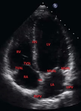
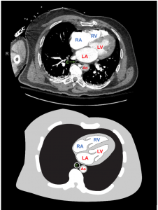
Parasternal Long Axis View In Normal Echocardiogram Echocardiogram Diagnostic Medical Sonography Cardiac Sonography

Pulmonary Hypertension Radiology Reference Article Radiopaedia Org
Ischemic congenital valvular heart.
Rv lv ratio radiopaedia.

Acute Reversible Pulmonary Hypertension And Right Heart Failure From Cocaine Toxicity Radiology Case Radiopaedia Org

Asymmetric Septal Hypertrophic Cardiomyopathy Systolic Anterior Motion Of Mitral Leaflet Radiology Case Radiopaedia Org

Optimal Threshold For The Diagnosis Of Anemia Severity On Unenhanced Thoracic Ct A Preliminary Study European Journal Of Radiology

Image Result For Parasternal Long Axis View Echocardiogram Ultrasound Future Doctor

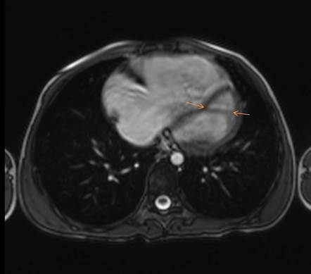
Rvot And Pa Mri Thoracic Cardiac

Extensive Acute Pulmonary Emboli With Right Heart Strain Radiology Case Radiopaedia Org

Link To And Excerpts From Pocus Cases 1 Pulmonary Embolism And Right Heart Strain From Emc Tom Wade Md
Left Ventricular False Tendon Radiology Reference Article Radiopaedia Org

Apical Hypertrophic Cardiomyopathy A Lv Angiography Demonstrates Apical Hypertrophy Hypertrophic Cardiomyopathy Human Body Anatomy Diastolic Heart Failure

Mass In Right Atrium Echocardiogram Hospital Humor Cardiology

Apical Four Chamber View Echocardiogram Ultrasound Sonography

Pulmonary Embolism Radiology Reference Article Radiopaedia Org

Type B Aortic Dissection Transesophageal Echocardiogram Tee Youtube Aortic Dissection Echocardiogram Cardiac Sonography

Apical 5 Chamber Cardiac Sonography Diagnostic Medical Sonography Echocardiogram
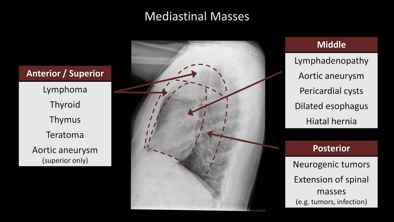
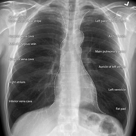
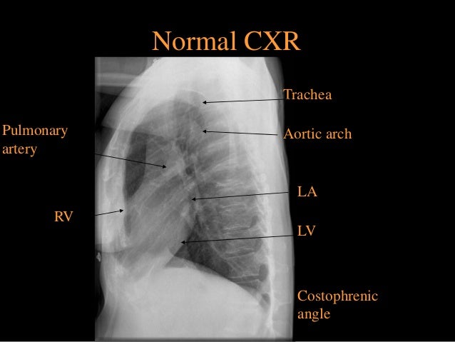
How To Interpret A Chest X Ray Lesson 5 Cardiac Silhouette And Mediastinum Youtube

Hypertrophic Cardiomyopathy Radiology Reference Article Radiopaedia Org
Radiology Chest Imaging

C Transposition Of The Great Arteries The Four Chamber View Was Normal The Great Arte Diagnostic Medical Sonography Cardiac Sonography Medical Ultrasound

Quantification In Pediatric Echocardiography Pediatrics Youtube Cardiology
Https Pubs Rsna Org Doi Pdf 10 1148 Rg 284075031

Left Ventricular Non Compaction Radiology Case Radiopaedia Org

Athlete Heart Syndrome Radiology Reference Article Radiopaedia Org
Em 3am Pacemaker Aicd Complications A 69 Year Old Female Presents
Http Med Mu Com Wp Content Uploads 2018 08 Radiology At A Glance Chowdhury Rajat Wilson Iain Pdf
Source : pinterest.com
